

Intraoperative monitoring of manifestations of reversible and irreversible myocardial ischemia/reperfusion injury during heart surgery, both without (off pump) and with cardiopulmonary bypass and cardioplegia, is important for optimizing the tactics of surgical intervention and cardiac anesthesia support. Timely detection of the onset of myocardial ischemia during surgery allows the cardiac surgeon to eliminate its cause, thus improving the immediate and long-term results of the surgery. The scientists of the Myocardial Microcirculation and Metabolism Department and Nanotechnology Research Laboratory of the Experimental Medicine Institute at Almazov Centre, together with the specialists of the Laser Centre at Pavlov State Medical University, are making scientific advancements in the field of rapid detection of perioperative ischemia. The specialists of the Laser Centre have extensive experience and a number of joint publications on autofluorescence organoscopy in ischemia/reperfusion injury of various organs. The jointly developed FOS-2 spectrofluorimeter is based on NADH fluorescence and is capable of detecting tissue ischemia in the first minutes. The spectrofluorimeter was designed at Pavlov State Medical University and Almazov Centre based on the previous developments.
The specialists of Almazov Centre showed that the fluorophore indocyanine green (ICG) accumulates in the area of myocardial infarction, and the accumulation area becomes visible in infrared light 15–20 minutes after its intravenous administration if the blood flow in the infarction-associated coronary artery is restored.
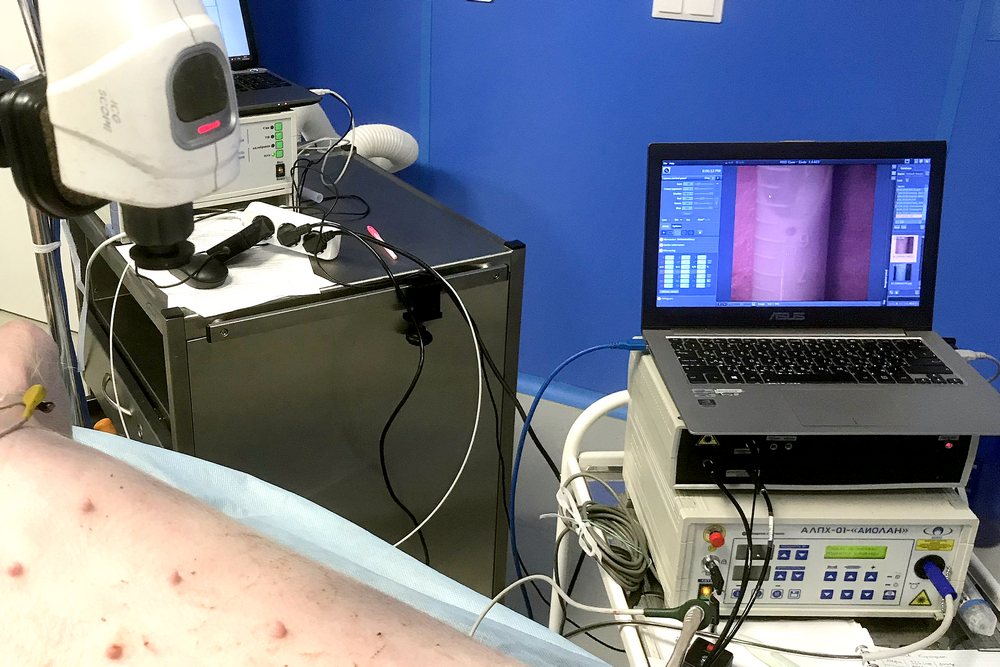
ICG is used in surgery and ophthalmology for fluorescence angiography. The fluorescent image is recorded upon excitation of the studied area at 780–810 nm, with registration at 820–900 nm. In this study, a recording unit with a special ICG-scope camera was used, capable of displaying the image in the visible and infrared spectra. After modeling myocardial ischemia by temporary ligation of the coronary artery, the animals were administered intravenous ICG both during the restoration of blood flow and at various time intervals, up to 24 hours, and after 30 minutes, ICG fluorescence was recorded from the surface of the organ and from its sections.
 |
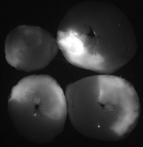 |
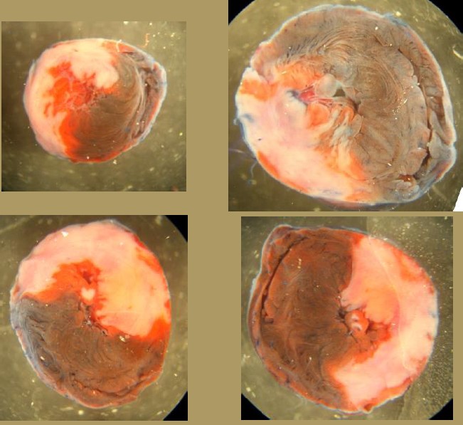
| Myocardial infarction | ICG fluorescence |
Imaging of the sections of the hearts in rats showed that the intensity of ICG fluorescence in the infarction area decreases one day after the ischemia/reperfusion injury. This phenomenon is not specific for rats and has been modeled in pigs. The developed technology complements the available diagnostic methods and can be used to determine the location and size of myocardial infarction from the surface of the heart. This opens up the possibility of using it for rapid intraoperative diagnosis of myocardial infarction both during cardiac surgery in a patient and in experiments on animals. The results obtained in experiments on rats and pigs make it possible to recommend recording the fluorescence intensity of ICG for intraoperative diagnosis of ischemia/reperfusion injury. An algorithm has been proposed to include the ICG fluorescence imaging into the existing techniques for monitoring the myocardium state during cardiac surgery.